
Clinical reports & patient experience
For some people, Morgellons means a deep facial wound, a sore that repeatedly opens, or a scar that changes how they look and live. Fibers and unusual sensations may bring someone to the label, but the wound can become the most urgent problem: pain, lost tissue, difficulty eating or washing, and uncertainty about whether the skin will heal.
Published reports document substantial ulceration and disfigurement in people described as having Morgellons or related presentations. They also offer different explanations for the damage. A useful account needs to preserve both facts: the wounds deserve attention, and the label does not establish their cause.
What do Morgellons sores look like?
“Gaping,” “crater-like” and “holes in the skin” communicate something important about appearance. A clinical description can make those observations more precise.
An ulcer involves loss of the entire epidermis and at least part of the underlying dermis; it may extend into deeper tissue. An erosion involves partial or complete loss of the epidermis without loss of the dermis. The International League of Dermatological Societies recommends describing an ulcer’s depth, border, base and surrounding tissue. [1]
An erosion stays within the epidermis; an ulcer extends into the dermis. Layer depths are simplified and not to scale.
General teaching diagram, not a photograph or reconstruction of a reported case.
The distinction matters because an open ulcer, a crusted surface and a depressed scar can look similar in a photograph. A raised rim also needs to be distinguished from elevation of the whole lesion.
A clinical example appears in Middelveen and colleagues’ 2021 case report. Figure 1 describes the patient’s back lesions as “ulcerated, punctate erosions with ragged edges,” which the authors call typical of Morgellons. This is the description supplied by a research group proposing an infectious explanation. It describes ragged wound edges, rather than establishing a characteristic raised rim. [2]
The caption also combines ulcer and erosion terminology, so its wording alone cannot resolve the depth distinction set out in the ILDS definitions. [1,2]
Published cases of deep wounds and disfigurement
The following reports use different definitions of Morgellons. Some describe patients within a proposed infectious-disease model; others use a delusional-infestation framework or arrive at a different diagnosis. They should not be combined into a uniformly diagnosed patient group.
Facial, scalp and oral wounds
| Report | Wound or complication described | Interpretation and limits |
|---|---|---|
| Caputo et al., 2022 [3] | A woman with substantial ulceration and scarring involving the face, trunk and arms. | The abstract reports fibers after multiple biopsies, no evidence of Borrelia infection, and a diagnosis in the setting of delusional infestation. Antipsychotic treatment began, but follow-up was lost. Healing was not documented. |
| Middelveen et al., 2020: patient M-13 [4] | Deep facial ulcerations, mucosal ulcers and secondary Staphylococcus aureus infection. | One patient in a selected 16-patient cohort framed by the authors as Borrelia-associated disease. The account documents severe wounds without establishing their frequency or a cause shared by every similar lesion. |
| Fiorese et al., 2025 [5] | A 54-year-old woman with scalp lesions described as ranging from small defects to deep, scooped-out wounds several centimeters across. | The authors described intensely itchy follicular papules, repeated scratching and a compulsive component. The text describes deep defects, whereas Figure 1 is captioned as superficial excoriations. |
| Alhendi and Burahmah, 2023 [6] | Oral and perioral ulceration extending onto facial skin, followed by scarring and hypopigmentation. | The patient reported using a needle and razor to remove perceived organisms. The authors diagnosed primary delusional parasitosis and reported improvement after local treatment and psychiatric care. |
Earlier coverage: I discussed Caputo et al.’s case in my July 2022 commentary on the woman with disfiguring cheek wounds on The New Morgellons Movement. That article emphasizes possible infection and additional testing. The case report’s accessible abstract reports no evidence of Borrelia infection and no follow-up outcome; it does not establish an infectious cause. [3]
These are distinct clinical accounts. A patient’s report that an opening appeared before touching it belongs in the history. A report of injury with a tool belongs there too. Neither sequence should be assumed for another patient.
A chin ulcer ultimately diagnosed as trigeminal trophic syndrome
Chrispim Gimenez and colleagues described a 33-year-old woman with a painful chin lesion present for 1.5 years. She reported material emerging from it that was not visible on examination. Her history included pituitary-tumor resection, although no clear preceding neurologic injury was identified. [7]
The authors diagnosed trigeminal trophic syndrome from the presentation and response to mental nerve blocks. They attributed ulceration to manipulation associated with altered sensation in the nerve’s territory. Pain improved after the first block, followed by reepithelialization. She also received pregabalin, droperidol and valproic acid during the initial encounter; later blocks followed, and pregabalin continued as needed. This course does not isolate the effect of a single treatment. [7]
Facial scars and damage around the eye
Robles and colleagues described a contracted, disfiguring scar between the eyebrows after prior ulceration in one of three patients reporting fibers. The publisher’s figure caption attributes the scarring to manipulation intended to extract fibers; the authors discuss the cases as delusions of parasitosis. This account is limited to the abstract and accessible case and caption excerpts. [8]
Sandhu and Steele reported a woman with self-reported Morgellons, an outward-turning lower eyelid, broken eyelashes and an ulcerated upper-eyelid lesion initially concerning for carcinoma. Biopsy findings were consistent with excoriation. Repair was deferred because of concern that continued excoriation would disrupt the surgical wound. [9]
Ma and colleagues’ 2019 report describes corneal perforation and surgical removal of an eye under the Morgellons label. Only its title and bibliographic record were available for this review; they do not support reconstructing the intervening clinical sequence or assigning a mechanism here. [10]
Reconstructive surgery after extensive injury
Nunziato and colleagues’ four-patient series included a man with bilateral elbow pterygia—skin webs restricting movement—treated with releases, flaps and grafts. The authors attributed injury to self-excoriation, reportedly mainly during sleep. Cases came from an informal hand-surgeon survey. [11]
Savely’s photographic sequence
Ginger Savely’s book gallery provides a separate form of documentation. Images 12–16 follow the same male patient through deep circular facial wounds, worsening, temporary healing and openings at new sites. [12]
The sequence illustrates recurrent, disfiguring wounds in a patient Savely identifies as having Morgellons. It is clinician-published photographic documentation rather than a peer-reviewed report containing a complete diagnostic investigation. The captions do not establish why the wounds formed or which treatment produced improvement.
View the original photographs and captions. The gallery contains graphic clinical images. Looking at the sequence matters: temporary closure and later recurrence tell a different story from a single before-and-after pair.
What these reports say about tissue damage
Six of the nine peer-reviewed reports in the wound-case section above attribute damage at least partly to scratching, excoriation or other manipulation: Fiorese, Alhendi, Chrispim Gimenez, Robles, Sandhu and Nunziato. [5–9,11]
The evidence behind those attributions varies. Alhendi and Nunziato include patients’ accounts; Sandhu reports a biopsy consistent with excoriation; Chrispim Gimenez combines clinical interpretation with treatment response; Fiorese describes repeated scratching with a compulsive component; and Robles attributes scarring to manipulation. A diagnostic label alone carries different information from a reported act, an examination finding or a treatment response.
The other three reports have different limits: Middelveen frames M-13 within a Borrelia-associated model; Caputo’s accessible abstract supplies a delusional-infestation framework without detailing the wound mechanism; and Ma’s mechanism remains unverified here. [3,4,10] Nunziato’s broader mass-psychogenic-illness framing should likewise be distinguished from the individual observations. [11]
Selection matters in both directions. Clinical setting, case definition and publication choices affect which patients appear in psychiatric reports and infection-oriented cohorts. Counting explanations in this selected literature cannot tell us how often each mechanism occurs among everyone using the Morgellons label.
What the cases establish: Severe wounds are documented, and most of these reports attribute damage at least partly to manipulation, on differing evidence. They do not establish a single cause or the population frequency of deep wounds. Recognizing a contribution from manipulation in a particular case does not diminish the injury or remove the need to investigate pain, altered sensation, infection and impaired healing.
Raised edges, pigment changes and thick scars
The Morgellons-specific sources reviewed here provide sparse evidence for a consistent raised rim around an ulcer. They describe several different structures, which should be recorded separately.
Raised and rolled edges are shown in profile; ragged is a surface outline. Undermining extends beneath the visible margin.
These features describe appearance, not cause. General schematic; not to scale.
Alongside the ragged-edged back lesions described above, Middelveen’s 2021 report shows expanding, ring-shaped rashes with “a raised, advancing, erythematous border.” The authors interpreted these as secondary erythema migrans. Figure 2’s raised border surrounds a rash; it is not a description of the rim of the wounds in Figure 1. Neither caption establishes a measured wound depth. [2]
Healing changes are another category. In a 2006 practitioner letter, Randy Wymore and Rhonda Casey described poorly healing lesions followed by “unusually thick, membranous scarring upon eventual healing.” The letter was hosted by the Morgellons Research Foundation. [13] Savely and Stricker’s selected 122-patient cohort separately listed hyperpigmented scars after healing among commonly reported symptoms. [14]
At the fifth annual Morgellons conference in 2012, Peter Mayne described chronic elevated papules and plaques, with eschars and ulceration in more advanced lesions. He also discussed post-inflammatory hyperpigmentation and hypopigmentation and linked lichenification—skin thickening with accentuated markings—to continual scratching. Mayne later coauthored Middelveen’s 2013 filament study, linking this conference account to the same research group advancing an infectious model of Morgellons. [15]
For documenting a wound, this article recommends distinguishing:
- Elevation: Is the whole lesion raised, or only its border?
- Surface: Is the center open, crusted, plugged or healed?
- Symptoms: Is it painful, itchy, tender or numb? Did that change before or after the skin opened?
- Pigment: Is the skin darker or lighter than its previous appearance?
- Scarring: Is there thickened, contracted or depressed healing tissue?
- Sequence: Which change appeared first, and how quickly did it progress?
These observations preserve information that a general description such as “rash” can lose.
All four profiles show closed surfaces. Dashed lines in the keloid panel mark the original wound limits.
General scar profiles, not reconstructions of the patients described here. Not to scale.
Other conditions that can produce ulcers or crater-like lesions
A raised edge and depressed center can occur in conditions with very different mechanisms. Other disorders produce open wounds through persistent itch or repeated injury. Both kinds of comparison matter here.
The following are clinical comparisons, not diagnoses retrospectively assigned to the Morgellons cases above or a ranking of their likelihood. Shape, pain, itch, progression and exposure history help guide assessment; none identifies or excludes a diagnosis by itself.
| Condition | Appearance, symptoms and progression | What the comparison helps distinguish |
|---|---|---|
| Basal cell carcinoma [16] | A slowly growing lesion that may bleed or ulcerate. Nodular facial BCC can have a pearly surface and a central depression with a rolled edge. | A persistent facial ulcer with a raised border needs assessment for a skin tumour as well as inflammatory or infectious causes. |
| Squamous cell carcinoma [17] | An enlarging, scaly or crusted lump that may ulcerate over weeks to months; often tender or painful. | Pain does not establish infection. In Sandhu’s eyelid case above, carcinoma was an initial concern before biopsy supported excoriation. [9] |
| Keratoacanthoma [18] | A rapidly growing, firm nodule with a central keratin plug, producing a crater-like profile. | A plugged tumour differs from an open ulcer. Clinical appearance may not reliably distinguish keratoacanthoma from SCC. |
| Prurigo nodularis [19] | Intensely itchy, firm nodules. Scratching can thicken and enlarge lesions; some become ulcerated and leave scars or pigment changes. | This is a chronic itchy skin disorder. An itch–scratch cycle does not by itself imply deliberate injury or establish a psychiatric diagnosis. |
| Dermatitis artefacta [20] | Self-induced lesions with variable appearances, sometimes sharply outlined, linear or geometric, often on accessible skin. | The mechanism requires assessment. This diagnosis is not a synonym for all scratching, and a psychiatric history alone does not establish it. |
| Pyoderma gangrenosum [21] | A rapidly enlarging, often very painful ulcer with a purple, undermined edge—tissue loss extending beneath the visible margin. | This inflammatory condition can worsen after injury. It requires assessment that considers infection and other mimics. |
| Ecthyma [22] | A firm crust over an inflamed, hardened ulcer, sometimes with drainage; healing may leave a scar. | This deeper bacterial infection needs to be distinguished from secondary infection of a wound that began another way. |
| Cutaneous leishmaniasis [23] | Lesions may develop raised borders and central craters, sometimes covered by crust. They are usually painless, but bacterial superinfection can make them painful. | Relevant geographic exposure and laboratory detection of parasites or their DNA matter. CDC cautions that clinical signs alone are insufficient to distinguish it from other conditions. |
| Syphilitic gumma [24] | A destructive lesion with central tissue loss. The facial case discussed below was painless and had a raised red border. | Appearance alone cannot identify syphilis; history, tissue findings and serology must be interpreted together. |
The useful question is therefore more specific than whether a sore resembles a crater: what produced the elevation, what happened to the central tissue, and what findings support the proposed explanation?
A diagnostic correction can run in either direction
Kłosowicz and colleagues reported a 47-year-old woman with paranoid schizophrenia and a painless, deep facial ulcer. The authors linked their provisional dermatitis artefacta diagnosis to an unclear psychiatric history and Enterococcus faecalis in tissue culture. The lesion progressed despite antibiotics. Biopsy findings and a reactive treponemal test (TPHA), with a negative RPR, led them to suggest gummatous syphilis. They subsequently learned that she had been diagnosed with syphilis during pregnancy about 25 years earlier and treated with injections; further treatment details were unavailable. Penicillin and local wound care were followed by gradual healing with scarring. [24]
That history matters because treponemal tests can remain reactive after treatment. [25] The serology therefore needed to be considered alongside the lesion, biopsy and prior-treatment history; it did not, by itself, establish the ulcer’s cause. [24,25]
This was not a Morgellons case. It illustrates two limits: psychiatric history cannot substitute for evidence of how a wound formed, and an organism cultured from a wound does not necessarily identify its initiating cause.
Read it alongside the trigeminal trophic syndrome case, where the Morgellons presentation gave way to an explanation involving altered sensation and manipulation. [7] Nunziato’s child case, discussed below, shows another hazard: a Morgellons attribution can obstruct treatment of an infected wound. [11] Each case calls for examining the evidence behind the initial explanation and revising it when warranted.
Media context: a vivid description of sores
The language used in public discussion has a separate history. A fan transcript of the 2011 Ancient Aliens episode “Aliens, Plagues and Epidemics” attributes this description to Cliff Mickelson, credited as a Morgellons patient:
“The overt dermatological symptoms would be open sores on the skin—usually round, volcanic almost looking in nature.” [26]
This passage shows how an account of visible wounds appeared in a television programme built around extraterrestrial speculation. It is included as media context, not as clinical evidence. The metaphor conveys appearance; it cannot establish a characteristic lesion, a shared cause or a connection to any condition in the comparison table.
Document the wound and follow its healing
A useful consultation starts with a description of the wound’s location, dimensions, depth, border, base and surrounding skin. [1] Add a record of pain, itch, numbness, bleeding and drainage. Photographs help track appearance; they do not replace examination.
Bring a short timeline showing when the spot first appeared, whether it began as a bump or an opening, and what happened before it enlarged. Include treatments, products, dressings and any scratching or attempts to remove material. Note effects on sleep, eating, washing, work and social contact. Our photography guide explains how to preserve useful photographic context.
Video: how clinicians assess a wound
Protection, symptoms and recorded outcomes
In Nunziato’s series, finger excoriations healed after three weeks in a mitten cast. Another patient left grafts intact but developed leg excoriations once his elbows moved freely. [11]
These observations support discussing protection from further injury, including scratching during sleep when relevant. [11] Casts require clinical supervision. Ask which dressings or barriers suit the wound and how pain, itch or altered sensation will be managed. Improvement with protection does not by itself establish what initiated a lesion.
The 2020 M-13 account explicitly describes S. aureus infection as secondary. An organism found in an ulcer may represent an additional problem requiring treatment without identifying the process that first opened the skin. [4]
Follow-up should record whether a wound closes, remains closed, becomes less painful and permits ordinary activities again. Changes in distress or sensations also matter, but a prescription or referral alone is not a recorded healing outcome.
Further viewing: Wound Healing — Paul Liu, MD (8 minutes 45 seconds). In this 2022 interview, Liu discusses diagnostic mistakes, the burden of nonhealing wounds and research developments, including experimental work in mice. A transcript accompanies the video.
Do not let the label delay urgent assessment
Attributing an infected wound to Morgellons can delay care. Nunziato’s child patient had an infected finger after a cactus-thorn injury. Her mother refused recommended debridement, attributing the problem to Morgellons; staff involved Child Protective Services to secure care. Later joint fusion was reported, but the authors did not establish that delay caused the joint damage. [11]
Painful, hot, swollen skin needs urgent medical assessment. Suspected infection accompanied by confusion, faintness, rapid breathing or severe illness warrants emergency care. [27] Severe eye pain or a change in vision after eye injury also warrants emergency assessment. [28]
Questions to leave the appointment with answered
For someone living with a deep sore, the immediate goal is an assessment and a plan that address the wound itself. Useful questions include:
- What kind of lesion is this—an ulcer, erosion, crusted lesion, nodule or scar?
- What findings support the proposed explanation, and what alternatives remain?
- Does the border, depth or progression call for further investigation?
- How will pain, altered sensation, infection and further injury be addressed?
- When will healing be reassessed, and what change should prompt earlier review?
Sources and scope
This is a selective review of wound reports, descriptive sources and clinical comparisons, not a systematic review or an estimate of prevalence. Television interviews, conference material and clinician-published photographs are distinguished from journal reports. The nine-report synthesis concerns the wound-case section, not every source cited in this article. Access limitations are noted below.
Jeremy Murphree is the founder of Morgellons Survey and a patient advocate. AI assisted preparation of this draft. The article has not been reviewed by a clinician.
[1] Nast A, Griffiths CEM, Hay R, Sterry W, Bolognia JL. The 2016 International League of Dermatological Societies’ revised glossary for the description of cutaneous lesions. Br J Dermatol. 2016;174(6):1351–1358. doi:10.1111/bjd.14419. Glossary PDF. ↩
[2] Middelveen MJ, Haggblad JS, Lewis J, et al. Dermatological and Genital Manifestations of Lyme Disease Including Morgellons Disease. Clin Cosmet Investig Dermatol. 2021;14:425–436. doi:10.2147/CCID.S299526. Article. Figure 1 describes back lesions with ragged edges; Figure 2 describes raised borders around annular rashes. These are the authors’ descriptions, not independent measurements of the images. ↩
[3] Caputo V, Bonoldi E, Citterio A, Rongioletti F. The challenge of Morgellons disease: A patient with clinicopathologic correlation. Clin Dermatol. 2022;40(6):686–690. doi:10.1016/j.clindermatol.2022.07.003. PubMed. Abstract and accessible excerpts; full case and figures unavailable for this review. ↩
[4] Middelveen MJ, Martinez RM, Fesler MC, et al. Classification and Staging of Morgellons Disease: Lessons from Syphilis. Clin Cosmet Investig Dermatol. 2020;13:145–164. doi:10.2147/CCID.S239840. Article. See Results and Table 2, patient M-13. ↩
[5] Fiorese G, Caballero Uribe N, Trüeb RM. Morgellons-like Disease of the Scalp. Int J Trichology. 2025;17(2):133–136. doi:10.4103/ijt.ijt_72_24. Article. ↩
[6] Alhendi F, Burahmah A. Delusional Parasitosis or Morgellons Disease: A Case of an Overlap Syndrome. Case Rep Dent. 2023;2023:3268220. doi:10.1155/2023/3268220. Article. ↩
[7] Chrispim Gimenez M, Patel JR, DeAngelis J, Tausk F, Puelle MR. Trigeminal trophic syndrome presenting as Morgellons disease with a 2-year delay in diagnosis. JAAD Case Rep. 2026;68:77–79. Published online November 27, 2025. doi:10.1016/j.jdcr.2025.11.028. Article. ↩
[8] Robles DT, Olson JM, Combs H, Romm S, Kirby P. Morgellons disease and delusions of parasitosis. Am J Clin Dermatol. 2011;12(1):1–6. doi:10.2165/11533150-000000000-00000. Publisher. Abstract and accessible case/figure-caption excerpts; full article and figure not available for this review. ↩
[9] Sandhu RK, Steele EA. Morgellons Disease Presenting As an Eyelid Lesion. Ophthalmic Plast Reconstr Surg. 2016;32(4):e85–e87. doi:10.1097/IOP.0000000000000258. Author-institution abstract. Abstract-based account. ↩
[10] Ma J, Roelofs KA, Badilla J. Morgellons disease leading to corneal perforation and enucleation. Can J Ophthalmol. 2019;54(6):e285–e288. doi:10.1016/j.jcjo.2019.04.002. PubMed. Title and bibliographic record only; no abstract or full case available for this review. ↩
[11] Nunziato CA, Egeland BM, Gurman A, Henry SL. Morgellons Disease: The Spread of a Mass Psychogenic Illness via the Internet and Its Implications in Hand Surgery. HAND. 2021;16(6):NP5–NP9. doi:10.1177/1558944720976648. Article. ↩
[12] Savely G. Clinical photographs accompanying her 2016 Morgellons book, images 12–16. Clinician-published photographic sequence. Savely also coauthored reference 14. ↩
[13] Wymore RS, Casey R. Re: Morgellons Disease [letter to practitioners]. Oklahoma State University Center for Health Sciences. May 15, 2006. Archived letter (PDF). Wayback Machine capture: March 6, 2007. Practitioner correspondence originally hosted by the Morgellons Research Foundation; quotation checked directly in the archived PDF. ↩
[14] Savely VR, Stricker RB. Morgellons disease: Analysis of a population with clinically confirmed microscopic subcutaneous fibers of unknown etiology. Clin Cosmet Investig Dermatol. 2010;3:67–78. doi:10.2147/CCID.S9520. Article. Selected retrospective clinical cohort. ↩
[15] Mayne P. Presentation at the fifth annual Morgellons conference, 2012. Public conference abstracts, p. 4. Detailed observations and timestamps derive from a privately held recording at Morgellons Survey, 5Th Morgellons 2.mp4, duration 47:05; relevant passages 15:16–16:16 and 16:30–17:07. The recording is not publicly linked here. Mayne’s coauthorship is documented in Middelveen MJ, Mayne PJ, Kahn DG, Stricker RB. Characterization and evolution of dermal filaments from patients with Morgellons disease. Clin Cosmet Investig Dermatol. 2013;6:1–21. doi:10.2147/CCID.S39017. Article. ↩
[16] Oakley A; updated by Martin Fuentes A. Basal cell carcinoma. DermNet. Reviewed August 2025. ↩
[17] Oakley A. Cutaneous squamous cell carcinoma. DermNet. Updated December 2015. ↩
[18] Oakley A; updated by Tan K and Keefe M. Keratoacanthoma. DermNet. Updated November 2021. ↩
[19] Gonçalves RB. Nodular prurigo. DermNet. July 2022; reviewed February 2026. ↩
[20] Ngan V. Dermatitis artefacta. DermNet. 2003. ↩
[21] Oakley A; updated by Dhawan G. Pyoderma gangrenosum. DermNet. Updated March 2022. ↩
[22] Ngan V; updated by Gomez J. Ecthyma. DermNet. Updated January 2016. ↩
[23] Centers for Disease Control and Prevention. DPDx: Leishmaniasis. See also Abbott A, Chancey RJ, Roy SL. Leishmaniasis. CDC Yellow Book, 2026 edition; updated April 23, 2025. ↩
[24] Kłosowicz A, Pizun A, Pastuszczak M, et al. Large syphilitic gumma on the face mimicking dermatitis artefacta. Adv Dermatol Allergol. 2022;39(5):996–997. doi:10.5114/ada.2022.120892. Full text. ↩
[25] Centers for Disease Control and Prevention. Syphilis: STI Treatment Guidelines. See “Treponemal Tests and Reverse Sequence Algorithm.” Accessed October 21, 2026. ↩
[26] Mickelson C. Interview in Ancient Aliens: “Aliens, Plagues and Epidemics.” 2011. HISTORY programme listing identifies season 3, episode 7; Tubi listing. Quotation follows the fan transcript; it was manually verified by this author against episode audio. The cast listing credits Mickelson as a Morgellons patient. ↩
[27] NHS. Cellulitis. Reviewed April 18, 2024. ↩
[28] NHS. Eye injuries. Reviewed March 12, 2026. ↩
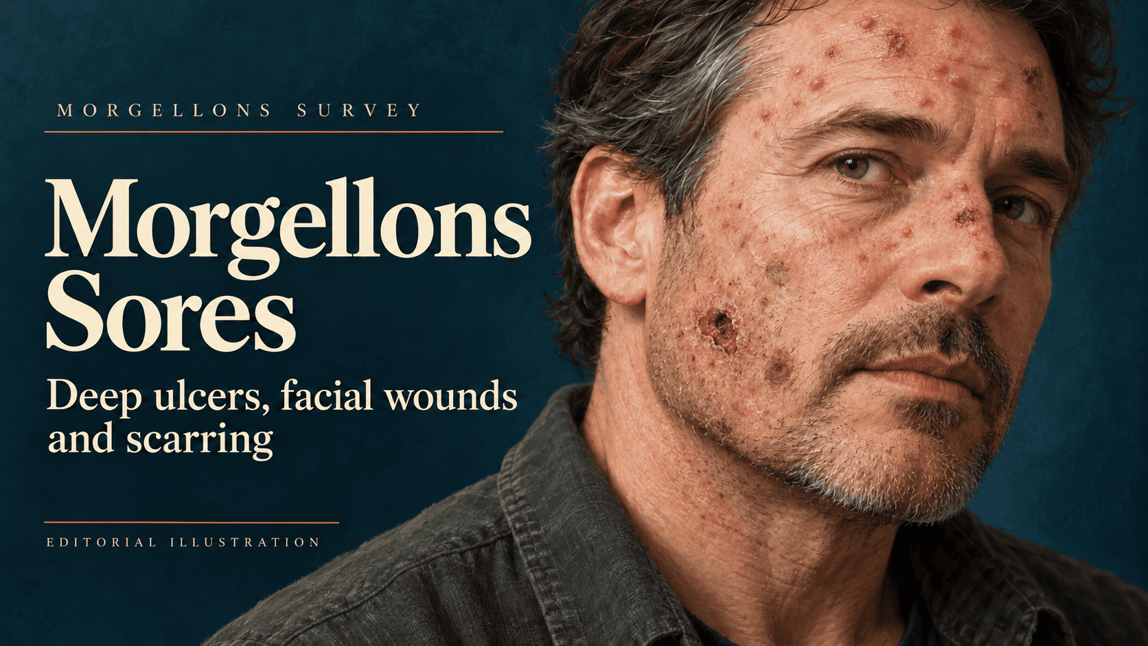
Featured illustration: AI-generated editorial illustration of a fictional person. It is not a clinical photograph or evidence from any case discussed above.